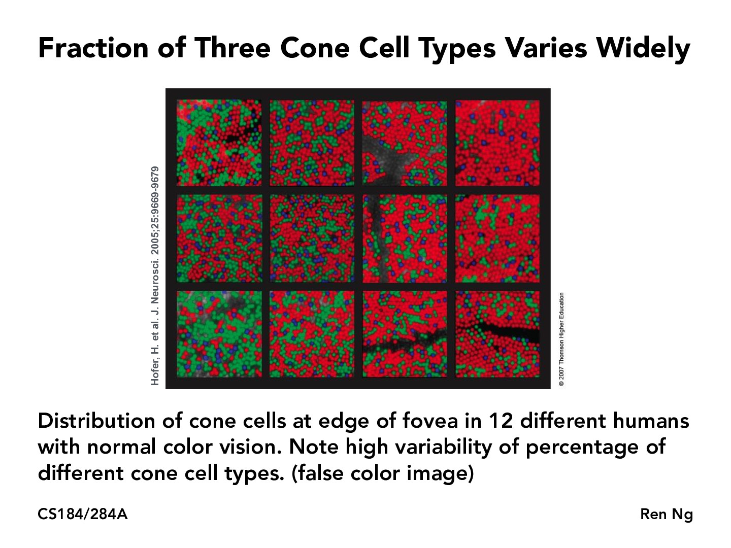
On the bottom left it seems like that person would perceive things to look more green while on the top right that person would perceive things to look more red. Is that actually what is the case or do these people perceive colors exactly the same?
Staffpsmanohar
These people perceive colors (more or less) exactly the same. By "perceive" I mean things that we can measure empirically, e.g. color matching experiments.
Also, while we typically label the L, M, and S cones with R, G, and B respectively, they don't really correspond to these colors.
knguyen0811
Are the empty spaces in the bottom-left and the grayscale dots in the middle-right supposed to represent no cones at all or non-functional ones, etc.?
iakopatran
Responding to the top comment, I think the confusion for me comes from these images depicting "normal" color vision but then saying how variability of different cone cell types can lead to false color image. To me I think normal color implies that their L, M, and S cones function properly according to the graph on slide 27. False color then is a result of the over activation of a certain cone (due to a higher concentration) leading to the perception of the false color. Just my guess though.
wangcynthia
Does color blindness relate to this somehow? Perhaps that people who have color blindness are deficient in a cone cell type?
jessicajyeh
Yes color blindness does relate to deficiencies in cone cells - according to this site, under the "causes" tab https://ghr.nlm.nih.gov/condition/color-vision-deficiency#genes , an absence in M or L cones affects red-green color vision, and impaired S cones alters perception of the color blue in particular (eg difficulty between shades of blue/green)
On the bottom left it seems like that person would perceive things to look more green while on the top right that person would perceive things to look more red. Is that actually what is the case or do these people perceive colors exactly the same?
These people perceive colors (more or less) exactly the same. By "perceive" I mean things that we can measure empirically, e.g. color matching experiments.
Also, while we typically label the L, M, and S cones with R, G, and B respectively, they don't really correspond to these colors.
Are the empty spaces in the bottom-left and the grayscale dots in the middle-right supposed to represent no cones at all or non-functional ones, etc.?
Responding to the top comment, I think the confusion for me comes from these images depicting "normal" color vision but then saying how variability of different cone cell types can lead to false color image. To me I think normal color implies that their L, M, and S cones function properly according to the graph on slide 27. False color then is a result of the over activation of a certain cone (due to a higher concentration) leading to the perception of the false color. Just my guess though.
Does color blindness relate to this somehow? Perhaps that people who have color blindness are deficient in a cone cell type?
Yes color blindness does relate to deficiencies in cone cells - according to this site, under the "causes" tab https://ghr.nlm.nih.gov/condition/color-vision-deficiency#genes , an absence in M or L cones affects red-green color vision, and impaired S cones alters perception of the color blue in particular (eg difficulty between shades of blue/green)